Anatomy Muscles Pelvis - Male Pelvis Skeleton Model With Ligaments Vessels Nerves Pelvic Floor Muscles Organs 7 Part 3b Smart Anatomy 1013282 3b Scientific H21 3 Genital And Pelvis Models Anatomical Models : Assoc prof craig hacking and dr tim luijkx et al.
byAdmin•
0
Anatomy Muscles Pelvis - Male Pelvis Skeleton Model With Ligaments Vessels Nerves Pelvic Floor Muscles Organs 7 Part 3b Smart Anatomy 1013282 3b Scientific H21 3 Genital And Pelvis Models Anatomical Models : Assoc prof craig hacking and dr tim luijkx et al.. Enclosing and protecting abdominopelvic and pelvic viscera. It is a broad flat muscle. The pelvis's frame is made up of the bones of the pelvis, which connect the axial skeleton to the femurs, and therefore acts in weight bearing of the upper body. Providing attachment for many muscles and ligaments used in locomotion; The pelvis is composed of the bony pelvis, pelvic floor, pelvic cavity, and the perineum.
The classification of the two groups under a common heading is. These muscles move the thigh toward the body's midline. Assoc prof craig hacking and dr tim luijkx et al. Large ligaments, tendons, and muscles around the hip joint hold the bones (ball and socket) in place and keep it from dislocating. Enclosing and protecting abdominopelvic and pelvic viscera.
Pelvic Floor Muscles Base For All Movement Anatomy from www.baselinehealing.com It is a broad flat muscle. Start studying anatomy and physiology of the pelvis. Assoc prof craig hacking and dr tim luijkx et al. Abdominal and pelvic anatomy encompasses the anatomy of all structures of the abdominal and pelvic cavities. It is usually divided into two separate anatomic regions: Learn vocabulary, terms, and more with flashcards, games, and other study tools. Continence, then pelvic muscle exercises may be effective. Cross the hip joint onto the thigh/leg 3.
Large ligaments, tendons, and muscles around the hip joint hold the bones (ball and socket) in place and keep it from dislocating.
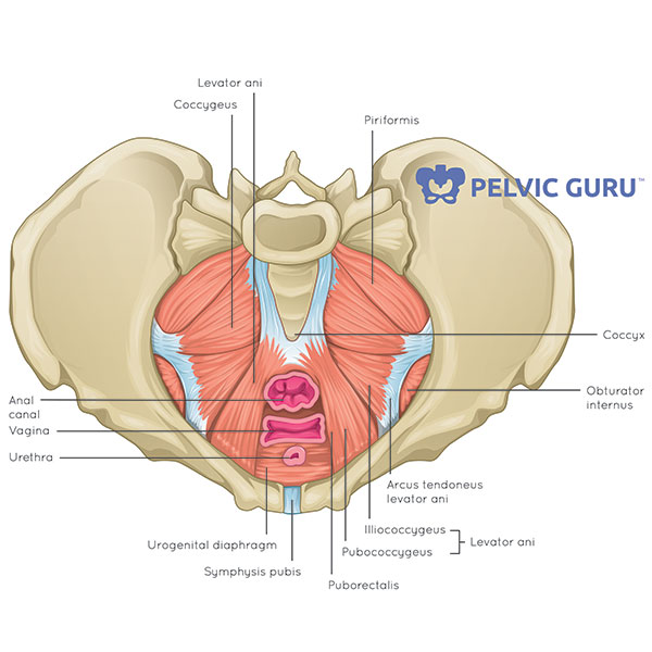
The levator ani muscles consist of three. Reviews the functional anatomy of the pelvic floor structures, the effects of age on urethral support and the urethral sphincter, and attempts to clarify Abdominal and pelvic anatomy encompasses the anatomy of all structures of the abdominal and pelvic cavities. To maintain the continence of urine and faeces. On each side, the pelvic diaphragm is formed by two most unequal muscles, the small coccygeus muscle behind, and the much larger and more important levator ani muscle in front. It separates pelvis from the perineum. These muscles move the thigh toward the body's midline. Cross the ls joint onto the trunk 2. As well as some basic images of disc pathology and stylised facet joint motion. Muscles that attach from the pelvis to the trunk and cross the lumbosacral joint muscles that attach from the pelvis to the thigh/leg and cross the hip joint pelvic floor muscles that are located wholly within the pelvis Enclosing and protecting abdominopelvic and pelvic viscera. The muscles of the pelvic floor are collectively referred to as the levator ani and coccygeus muscles. They form a large sheet of skeletal muscle that is thicker in some areas than in others.
The main function of the pelvic floor muscles are: The pelvis is composed of the bony pelvis, pelvic floor, pelvic cavity, and the perineum. The back of the abdomen consists of back muscles as well as the spine. Providing attachment for many muscles and ligaments used in locomotion; The muscles within the pelvis may be divided into two groups:
What Is The Pelvic Floor Your Pace Yoga from yourpaceyoga.com The muscles within the pelvis may be divided into two groups: On the other hand, if portions of those muscles are irretrievably lost, for example, due to complete. As well as some basic images of disc pathology and stylised facet joint motion. The muscles of the pelvic floor are collectively referred to as the levator ani and coccygeus muscles. It is a broad flat muscle. The muscles of the pelvis and hip control the vast range of movement of the legs and torso. On each side, the pelvic diaphragm is formed by two most unequal muscles, the small coccygeus muscle behind, and the much larger and more important levator ani muscle in front. Ct anatomy of the pelvis.
They form a large sheet of skeletal muscle that is thicker in some areas than in others.
These muscles move the thigh toward the body's midline. On the posterior side they are the glutei and on the anterior side the hip muscles extending into the thighs. On the other hand, if portions of those muscles are irretrievably lost, for example, due to complete. The muscles of the pelvic floor are collectively referred to as the levator ani and coccygeus muscles. The classification of the two groups under a common heading is. The main function of the pelvic floor muscles are: The levator ani muscles consist of three. Attached to the pelvis are muscles of the buttocks, the lower back, and the thighs. To maintain the continence of urine and faeces. Pelvic muscles ct anatomy and ct scan of the abdomen and pelvis shows a normal appendix 7 pelvic muscles ct anatomy pelvic muscles ct anatomy and ct scan of the abdomen and pelvis shows a normal appendix gallery at human diagram chart. The pelvis is composed of the bony pelvis, pelvic floor, pelvic cavity, and the perineum. Learn vocabulary, terms, and more with flashcards, games, and other study tools. Whenever someone talks about the pelvic floor in general, they are probably talking about these 5 muscles:
The muscles of the pelvis and hip control the vast range of movement of the legs and torso. Learn vocabulary, terms, and more with flashcards, games, and other study tools. Muscles that attach from the pelvis to the trunk and cross the lumbosacral joint muscles that attach from the pelvis to the thigh/leg and cross the hip joint pelvic floor muscles that are located wholly within the pelvis It takes origin from the inner aspect of pelvis along a line extending from the body of the pubis to the ischial spine. It is a basin shaped muscular diaphragm that helps to support the visceral contents.
Pelvic Floor Wikipedia from upload.wikimedia.org Psoas consists of a pair of deep muscles (psoas major and iliacus) located on each side of the pelvis in the abdomen. Large ligaments, tendons, and muscles around the hip joint hold the bones (ball and socket) in place and keep it from dislocating. It separates pelvis from the perineum. Reviews the functional anatomy of the pelvic floor structures, the effects of age on urethral support and the urethral sphincter, and attempts to clarify Use the mouse scroll wheel to move the images up and down alternatively use the tiny arrows (>>) on both side of the image to move the images.>>) on both side of the image to move the images. Attached to the pelvis are muscles of the buttocks, the lower back, and the thighs. Whenever someone talks about the pelvic floor in general, they are probably talking about these 5 muscles: The classification of the two groups under a common heading is.
Continence, then pelvic muscle exercises may be effective.
On each side, the pelvic diaphragm is formed by two most unequal muscles, the small coccygeus muscle behind, and the much larger and more important levator ani muscle in front. The levator ani muscles are the largest group of muscles in the pelvis. To maintain the continence of urine and faeces. Reviews the functional anatomy of the pelvic floor structures, the effects of age on urethral support and the urethral sphincter, and attempts to clarify Included in this group are the adductor longus, adductor brevis, adductor magnus, pectineus, and gracilis muscles. Describe the muscles of pelvic diaphragm. The greater pelvis is the space between the two wings of the ilium above the terminal line, while the lesser pelvis is the part of the pelvis below the terminal line. The right and left hip bones, plus the sacrum and the coccyx, together form the pelvis. Use the mouse scroll wheel to move the images up and down alternatively use the tiny arrows (>>) on both side of the image to move the images.>>) on both side of the image to move the images. Large ligaments, tendons, and muscles around the hip joint hold the bones (ball and socket) in place and keep it from dislocating. These muscles have attachments to the pelvis as follows: These muscles move the thigh toward the body's midline. Assoc prof craig hacking and dr tim luijkx et al.